Command Palette
Search for a command to run...
Polystyrene nanoplastics disrupt the intestinal microenvironment by altering bacteria-host interactions through extracellular vesicle-delivered microRNAs
Polystyrene nanoplastics disrupt the intestinal microenvironment by altering bacteria-host interactions through extracellular vesicle-delivered microRNAs
Wei-Hsuan Hsu You-Zuo Chen Yi-Ting Chiang Yi-Tsen Chang Yi-Wen Wang Kung-Ting Hsu Yi-Yun Hsu Pei-Ting Wu Bao-Hong Lee
Abstract
Nanoplastics (NP) are emerging environmental pollutants with potential risks to human health. This study investigates how polystyrene-NP exposure disrupts the intestinal microenvironment and barrier function through bacteriahost interactions. Using in vivo models and bacterial sorting technology, we show that NP accumulation in the mouse intestine alters the expression of intestinal miR-501-3p and miR-700-5p, compromising tight junction protein ZO-1 and mucin (MUC)−13 expression, thereby increasing intestinal permeability. NP increases miR-98-3p, miR-548z, miR-548h-3o, miR-548d-3p, miR 548az-5p, miR-12136, and miR-101-3p levels in extracellular vesicles (EVs) derived from goblet-like cells, which can interfere with ZO-1 expression. NP also induces gut microbiota dysbiosis, characterized by elevated Ruminococcaceae abundance and altered EV characteristics from goblet cells. Lachnospiraceae internalize NP, and their EVs suppress MUC-13 expression. These findings reveal a mechanism by which NP compromises intestinal integrity and indirectly alters intestinal microbiota composition, potentially leading to adverse health outcomes.
One-sentence Summary
National Cheng Kung University researchers demonstrate that polystyrene nanoplastics compromise intestinal barrier integrity by upregulating extracellular vesicle-delivered microRNAs (e.g., miR-501-3p, miR-98-3p) from goblet cells that suppress ZO-1 and MUC-13 expression, while inducing gut microbiota dysbiosis through altered bacterial extracellular vesicle characteristics, revealing a bacteria-host interaction mechanism underlying nanoplastic-induced intestinal permeability.
Key Contributions
- The study establishes an in vivo workflow for polystyrene nanoplastics in mice that combines oral nanoplastic administration, flow cytometric sorting of intestinal bacteria, fluorescence microscopy, and 16S sequencing to connect intestinal nanoplastic uptake with specific bacterial taxa and extracellular vesicle responses.
- The study demonstrates that polystyrene nanoplastic exposure disrupts the intestinal barrier by altering intestinal miR-501-3p and miR-700-5p expression, reducing ZO-1 and MUC-13 levels, increasing permeability, and inducing goblet-cell-derived EV miRNAs, including miR-98-3p, miR-548z, miR-548h-3o, miR-548d-3p, miR-548az-5p, miR-12136, and miR-101-3p, which can interfere with ZO-1 expression.
- The study identifies an EV-mediated host-microbiota pathway in which nanoplastics modify goblet-cell EV characteristics, elevate Ruminococcaceae abundance, promote Lachnospiraceae uptake of nanoplastics, and enable bacterial-derived EVs to suppress MUC-13 expression, as shown through gut microbiota profiling and differential taxa analysis.
Introduction
Nanoplastics are emerging environmental contaminants that can enter the intestinal tract after ingestion, making gut barrier integrity and host microbiome interactions a relevant exposure pathway. Prior studies have linked nanoplastic exposure to toxicity risk, but the mechanisms by which these particles disrupt host epithelial signaling and resident bacteria remain insufficiently resolved. In this work, the authors use mouse exposure models and bacterial sorting to show that polystyrene nanoplastics accumulate in the intestine, alter intestinal microRNA expression, reduce ZO-1 and MUC-13 levels, and increase permeability. They further show that extracellular vesicles from goblet-like cells and Lachnospiraceae-derived vesicles can suppress barrier-related proteins, while nanoplastic exposure reshapes microbiota composition. The authors contribute a bacterial-host mechanism linking nanoplastic exposure to weakened intestinal defense and potential health risk.
Dataset
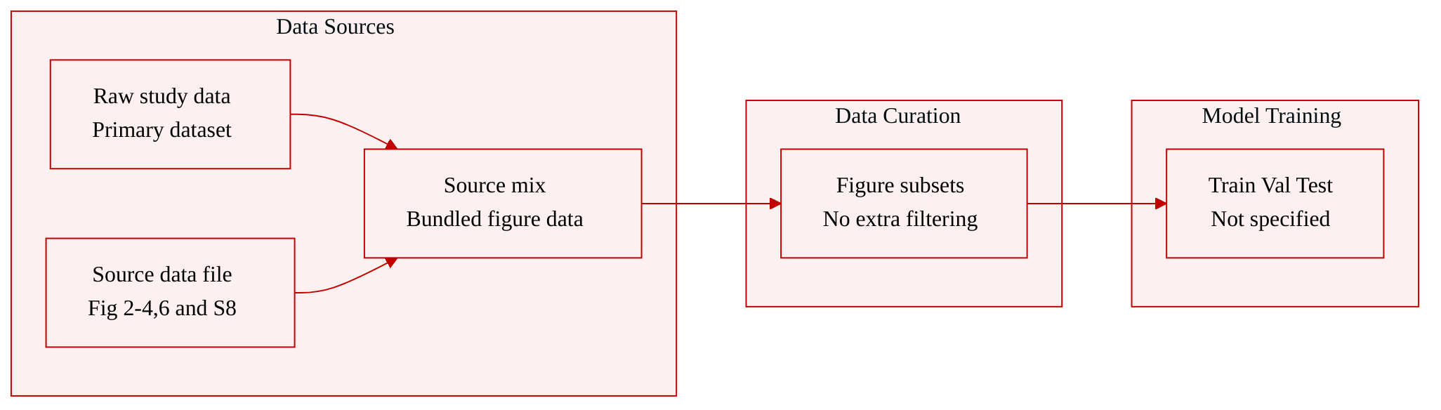
- The authors use raw data generated in the study as the primary dataset.
- Source data are provided with the paper and made available in the Supplementary Information and Source Data file.
- The included subsets are source data underlying Figures 2, 3, 4, and 6 and Supplementary Figure 8.
- The paper does not specify additional dataset splits, filtering rules, metadata schema, cropping strategy, or mixture ratios beyond providing the raw and source data associated with the reported figures.
Method
The experimental architecture of this study is organized as a multilayer pipeline that connects nanoparticle exposure to intestinal barrier dysfunction, host transcriptomic and microRNA regulatory changes, and downstream microbial communication assays. The workflow begins with physical characterization of nanoparticles, moves to controlled in vivo and in vitro exposure models, then applies histological, molecular, sequencing-based, and bioinformatic modules to map causal relationships between nanoparticle uptake, epithelial barrier damage, and microbiota-related signaling. The authors leverage an integrated design in which phenotypic observation guides molecular profiling, and predicted regulatory interactions are validated through RNA interference and protein-level assays.
The first module focuses on nanoparticle identity, dosing, and distribution. Nanoparticles are characterized by nanoparticle tracking analysis to verify particle count and size before exposure. In the animal model, fluorescently labeled nanoparticles are administered orally over a sustained period, after which colonic tissue, feces, serum, and other organs are collected. In parallel, cultured cell systems are used to examine cellular uptake under controlled concentrations and time points. Fluorescent nanoparticles can be imaged directly, allowing the authors to track intracellular accumulation and associate exposure levels with downstream molecular changes. This exposure framework supports both systemic barrier assessment in vivo and mechanistic testing in cellular models.
The barrier-function module links nanoparticle uptake to epithelial integrity. Enterocyte-like differentiated Caco-2 cells are used as an in vitro intestinal epithelial model, while murine colon tissue provides the in vivo counterpart. Tight-junction proteins, including ZO-1 and occludin, are assessed by immunofluorescence, immunocytochemistry, immunohistochemistry, and western blotting. Monolayer permeability is evaluated using an FITC-dextran flux assay, where increased translocation from the apical to basolateral compartment is used as a functional readout of barrier disruption. These assays are placed in direct relation to nanoparticle exposure, so that uptake, tight-junction protein depletion, and permeability increase are interpreted as a connected phenotypic chain rather than isolated measurements. Refer to the barrier assessment figure:
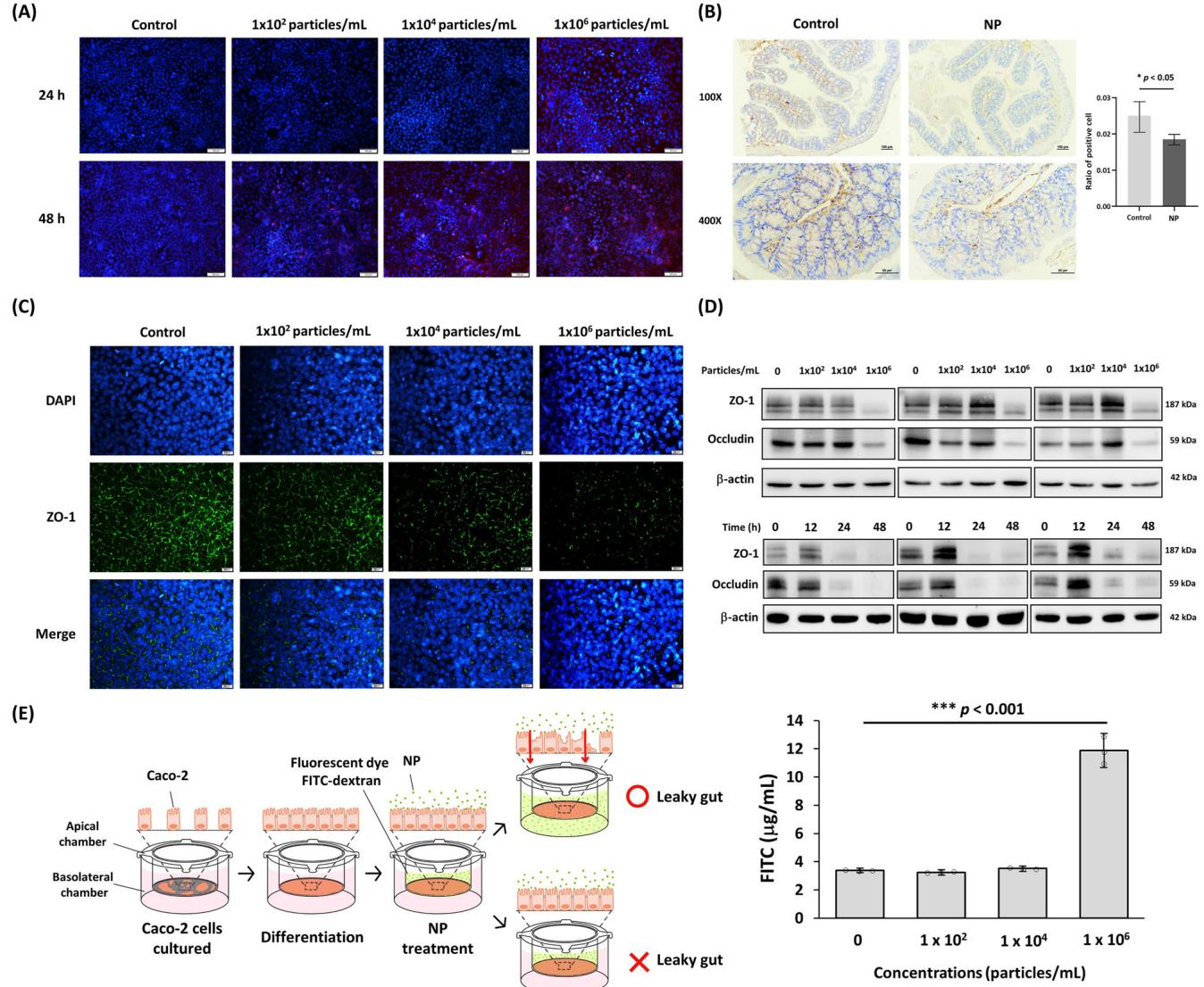
This module is designed to test whether nanoparticle exposure directly compromises epithelial barrier structure and function. The combined use of fluorescence imaging, tissue staining, protein quantification, and permeability flux creates an orthogonal validation strategy. The visual uptake patterns, ZO-1 staining changes, and FITC-dextran translocation data are integrated to confirm that exposure correlates with barrier loss across experimental scales.
The second major module addresses host molecular regulation through transcriptome and microRNA profiling. Total RNA is extracted from intestinal tissue or cells, processed into sequencing libraries, and subjected to quality trimming and differential expression analysis. Genes that meet thresholds of at least twofold change and statistical significance are retained for downstream interpretation. The authors apply principal component analysis and heatmap visualization to compare global expression patterns between exposed and control conditions. For microRNA profiling, small RNA sequencing data are processed through an established bioinformatic workflow, known microRNAs are quantified, and novel microRNAs are also considered during annotation. Differentially expressed microRNAs are selected using adjusted significance cutoffs and fold-change criteria, while target-gene prediction and pathway enrichment are used to infer biological consequences.
To convert candidate microRNAs into mechanistic hypotheses, the authors integrate prediction-based target searching with experimental validation. MicroRNA-target interactions are explored using target databases and hybrid-structure prediction, which estimates duplex stability and predicted binding regions. The selected candidate microRNAs are then introduced into differentiated epithelial cells through transient transfection, and the effect on target gene expression is evaluated by qPCR, immunofluorescence, and western blotting. Relative expression is commonly normalized using housekeeping genes and calculated through the 2−ΔΔCt framework, while protein abundance is normalized to loading controls. As shown in the miRNA regulatory figure:
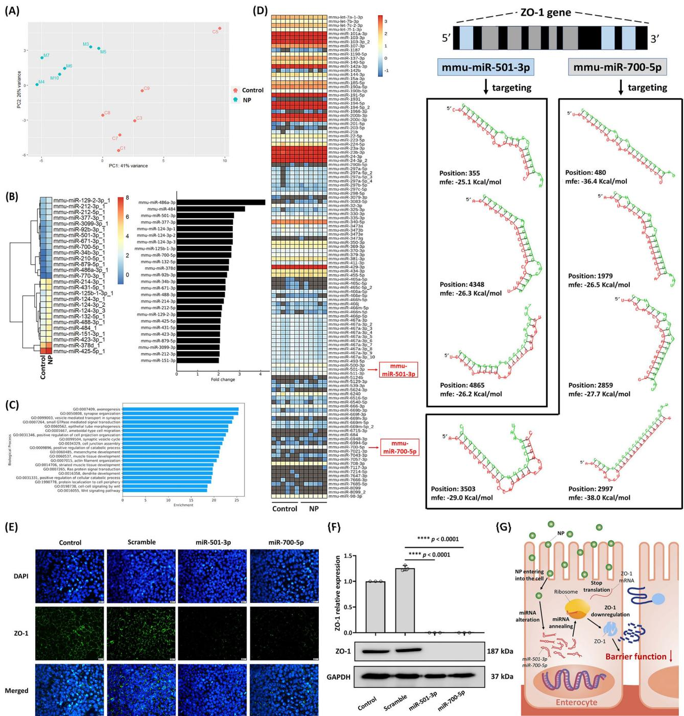
In this part of the pipeline, the authors connect microRNA alteration to tight-junction regulation. Rather than reporting only differential microRNA abundance, the workflow proceeds through target prediction, functional interference, and protein-level confirmation. This design allows the study to move from correlation to mechanism, particularly for microRNAs predicted to act on barrier-associated transcripts such as ZO-1.
A related mucosal barrier module extends the analysis from tight junctions to mucin expression and mucus layer integrity. MUC13 expression is evaluated across multiple systems, including differentiated enterocyte-like cells, goblet-like LS174T cells, and murine colon tissue. mRNA levels are measured by qPCR, protein levels by western blotting, localization by immunohistochemistry and immunocytochemistry, and mucus content by alcian blue periodic acid Schiff staining. MicroRNA targeting analysis is applied to MUC13 as well, with candidate microRNAs tested through transfection experiments. The goal of this module is to determine whether the same nanoparticle-induced regulatory state that affects tight junctions also suppresses mucosal protective factors. Refer to the mucosal barrier figure:
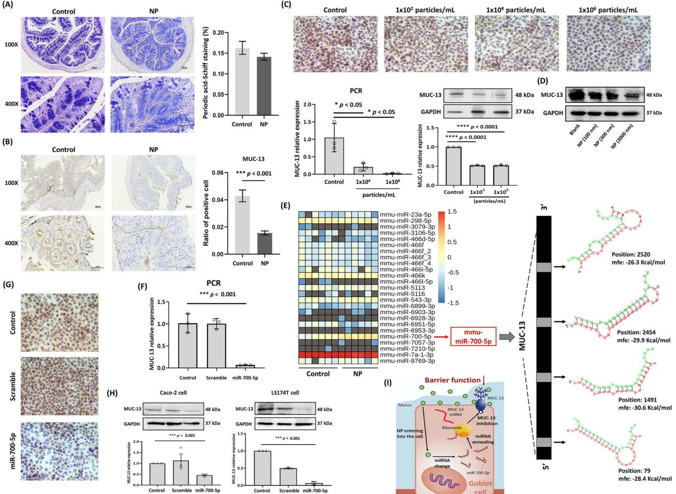
This portion of the experimental framework emphasizes convergence across readouts. Histological mucus staining, mucin protein abundance, mRNA expression, and microRNA perturbation are all used to test whether barrier degradation involves both junctional and secretory defenses. The inclusion of distinct cell models, goblet-like and enterocyte-like, allows the authors to separate cell-type specific effects and examine how mucin regulation may differ from tight-junction regulation.
The final component of the pipeline incorporates extracellular vesicle and microbial growth assays to examine possible cross-kingdom effects. Nanoparticle exposure is applied to microbial cultures, and extracellular vesicles are isolated from culture supernatants through sequential centrifugation, filtration, and resuspension steps. Vesicle particle number and size are assessed by nanoparticle tracking analysis. These vesicles are then applied to additional microbial systems, either alone or in combination with nanoparticle exposure, and growth responses are measured by optical density and direct enumeration. This module connects host barrier assays to microbial ecology by testing whether nanoparticle-associated vesicular signals can alter microbial growth behavior. In the broader experimental architecture, the microbial assay suite complements the host molecular modules by placing intestinal barrier changes within a larger model of host-microbe interaction.
Experiment
Using fluorescent polystyrene nanoplastics in mice and intestinal cell models, the study validated intestinal nanoplastic accumulation and evaluated barrier, mucin, and microbiota responses. Chronic oral exposure did not produce overt liver or kidney injury or obvious intestinal inflammation, but it was associated with increased body weight, impaired tight junction proteins, increased permeability, and reduced MUC-13-associated mucin expression. These effects were linked to altered host microRNA profiles and extracellular vesicle signaling, which indirectly promoted Ruminococcaceae expansion and Lachnospiraceae-related mucin suppression, ultimately driving gut microbiota imbalance.